PPT Dr. Altdorfer The tongue Anatomy, histology, innervation PowerPoint Presentation ID
In 33.3%, one of the terminal branches of the mylohyoid nerve after perforating the homonymous muscle, anastomoses with the lingual nerve in the lateral sulcus of the tongue (Sulcus lateralis linguae) achieving, in the author's opinion, the "mylohyoid or sublingual curl".

theanatomyofthetongue Google Search Anatomy Head, Anatomy Bones, Body Anatomy, Anatomy Of
Definition Der Sulcus lateralis linguae ist ein anatomischer Spaltraum, der sich zwischen dem Musculus mylohyoideus und dem Musculus hyoglossus befindet. Anatomie Der Sulcus lateralis linguae lässt sich als Verlängerung des Recessus sublingualis lateralis unterhalb der Mundschleimhaut verstehen. In ihm verlaufen folgende Strukturen:

Tongue muscles, Anatomy, Anatomy of the tongue
Surgical Anatomy of the Tongue Mahmoud F. Sakr Chapter First Online: 24 August 2022 609 Accesses Abstract The tongue (Latin, lingua; Greek, glos sa) is a unique organ located in the oral cavity. It has importance in the digestive system and is the primary organ of taste in the gustatory system.
Nose and tongue
Der Sulcus lateralis linguae (lat. für „seitliche Zungenfurche") ist ein zwischen dem Zungenboden und Unterkieferknochen verlaufender dreiseitiger Spalt bzw. Kanal. Er wird oben durch den Musculus hyoglossus, unten durch den Musculus mylohyoideus und seitlich durch den Unterkiefer begrenzt.

Human Anatomy Scientific Illustrations Nervus Lingualis HighRes Vector Graphic Getty Images
The Sylvian fissure, also known as the lateral sulcus or fissure, begins near the basal forebrain and extends to the lateral surface of the brain separating the frontal and parietal lobes superiorly from the temporal lobe inferiorly 3.The insula is located immediately deep to the Sylvian fissure.. Gross anatomy. The Sylvian fissure can be divided into superficial and deep portions 3,4.
TongueGross Anatomy & Applied Aspects. Dr.N.Mugunthan.M.S
Walls of Sulcus lateralis linguae-M. Mylohyoideus (lateral)-M. Hyoglossus (medial) Alveolo-lingual mucosa (superior) Sets found in the same folder. Anatomy Module#3 Part#1. 221 terms. anandam288. ANATOMY PART#2. 238 terms. anandam288. ANATOMY part#3. 179 terms. anandam288. Cranial nerves Midterm exam. 95 terms. anandam288. Other sets by this.
Terminal sulcus of tongue (Sulcus terminalis linguae) Kenhub
The sulcus lateralis linguae (Latin for "lateral tongue furrow") is a three-sided gap or canal running between the base of the tongue and the lower jaw bone. It is bounded above by the hyoglossus muscle, below by the mylohyoid muscle and laterally by the lower jaw.. The structures running in it are from top to bottom: Nervus lingualis; Sublingual vein (variable)

Dorsal Surface Of Tongue , Png Download Tongue Diagram Simple, Transparent Png , Transparent
In neuroanatomy, the lateral sulcus (also called Sylvian fissure, after Franciscus Sylvius, or lateral fissure) is one of the most prominent features of the human brain. The lateral sulcus is a deep fissure in each hemisphere that separates the frontal and parietal lobes from the temporal lobe.

Sulcus lateralis Ars Neurochirurgica
The lateral lingual groove (or sulcus) is a V-shaped space located between the hyoglossus and mylohyoid muscles. Figure 1. Lateral lingual groove Subscribe now to continue reading Join hundreds of successful students who use Meddists to ace their exams. Gain access to all of the material and topics, custom-made just for you. Continue
PPT Dr. Altdorfer The tongue Anatomy, histology, innervation PowerPoint Presentation ID
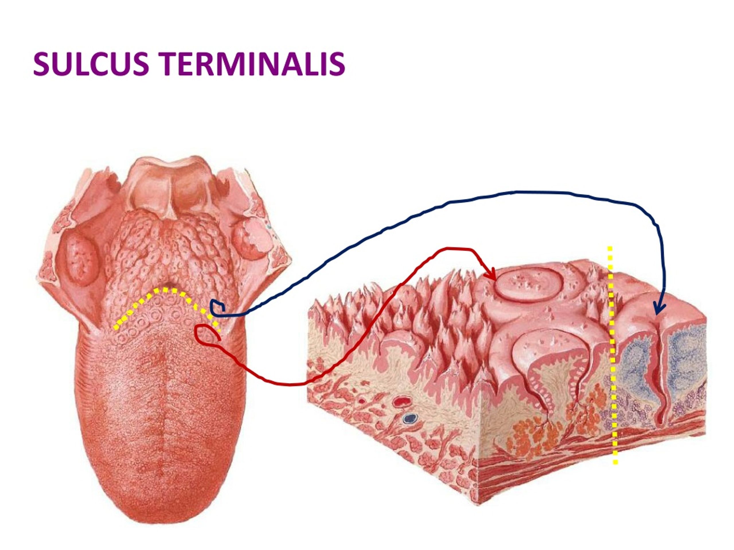
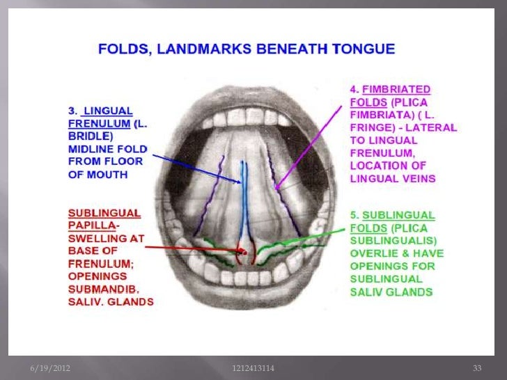
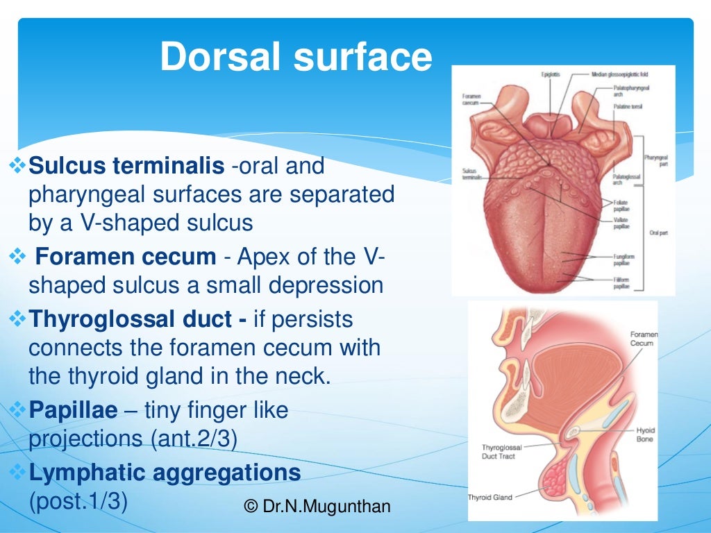
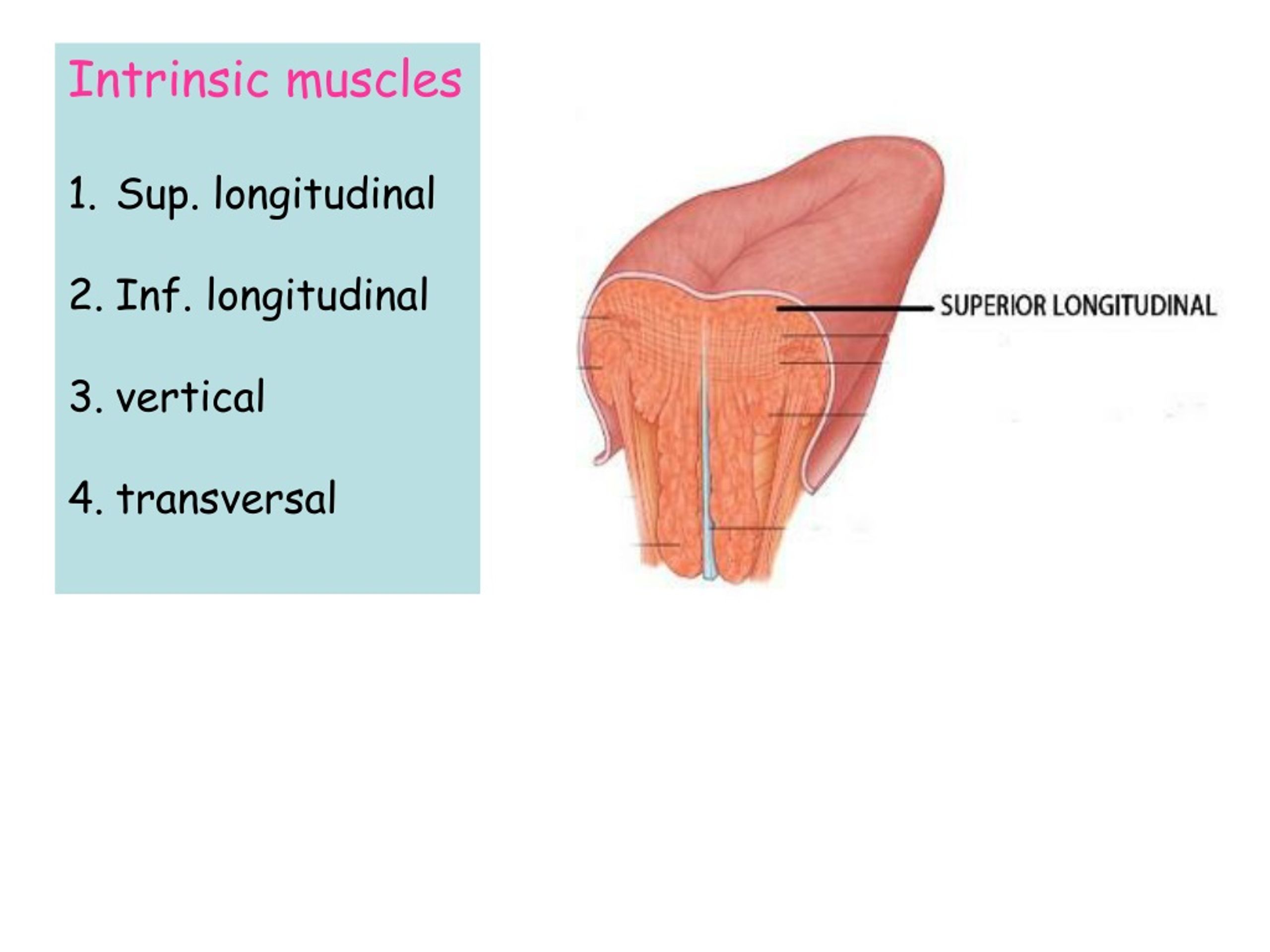
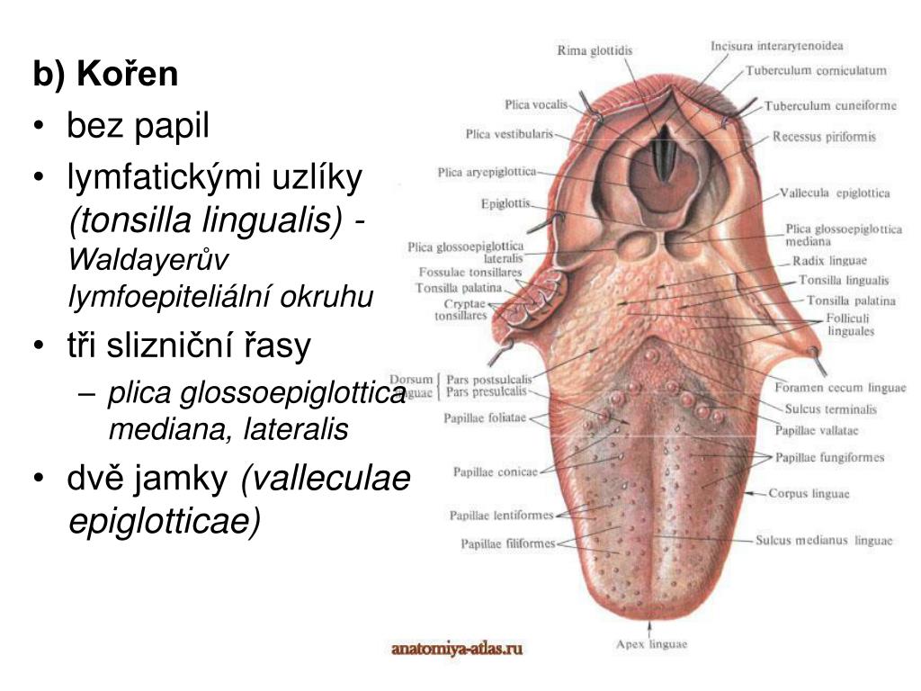
Dr. Altdorfer: The tongue - Anatomy, histology, innervation Sulcus terminalis Foramen cecum Root Follicular part Dorsum linguae Papillar part Apex linguae. Intrinsic muscles • Sup. longitudinal • Inf. longitudinal • vertical • transversal. SUBLINGUAL REGION • frenulumlinguae • deeplingualvein • sublingual fold • sublingualcaruncula (papilla)

Reference points used for measurements on the casts (A) lingual sulcus... Download Scientific
SULCUS LATERALIS LINGUAE SULCUS MEDIALIS LINGUAE Határai: medialis: m. hyoglossus lateralis: m. mylohyoideus felső: paralingualis nyálkahártya Tartalma: n. lingualis d. submandibularis n. hypoglossus + * v. comitans cum nervo hypoglosso.

TONGUE Anatomy for MBBS, NEET PG, AIIMS PG, FMGE & ALL PG YouTube
The lingual sulcus release technique described here finds its utility in squamous cell carcinoma of the tongue tumours in patients with limited mouth opening (grade I/II trismus), in which there is involvement of the ventral surface of the tongue with extension to the floor of the mouth (Fig. 1).Download : Download high-res image (315KB) Download : Download full-size image
Tongue Nerve and blood supply (lingual artery) Kenhub
The torus linguae, a raised portion of the tongue caudal to the lingual fissure. The buccal papillae, are conical papillae lining the inside of the cheeks. The surface of the tongue is covered in papillae, including the large round vallate papillae (with a moat encircling each one) near the root of the tongue.

PPT 23 PowerPoint Presentation, free download ID2246527
OBJECT: The sylvian fissure or lateral sulcus is the most identifiable feature of the superolateral brain surface and constitutes the main microneurosurgical corridor, given the high frequency of approachable intracranial lesions through this route.
PPT Trávicí systém (apparatus digestorius) PowerPoint Presentation ID4148661
When looking at the lateral surface of the cerebrum, we can recognize the lateral sulcus as a deep groove that separates the temporal lobe below from the frontal lobe above. This groove has a central part at the base of the brain, which becomes clearer when viewed from the lower side. From there, it extends sideways between the frontal and temporal lobes. As it reaches the outer surface of the.

Pin by Will Housell on Lickilicky Throat anatomy, Tongue, Anatomy
The lateral sulcus (sulcus lateralis of Sylvius), known for a long time as the Sylvian fissure, between the frontal and temporal lobes, has three branches: the anterior (ramus anterior) or horizontal ramus, the ascending (ramus ascendens) or vertical ramus and the posterior ramus (ramus posterior), separating the parietal and temporal lobes.